
Glaucoma is one of the leading causes of permanent blindness, yet most people who have it don’t know it yet. There are often no headaches, no pain, no blurry mornings to tip you off. The condition moves quietly, and by the time many patients notice something is wrong, real damage has already been done. Keep reading to understand how glaucoma quietly damages your vision, and what you can do about it.
The Part of Your Eye That Glaucoma Targets
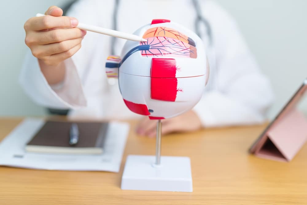
Every image you see travels from your eye to your brain through the optic nerve. The optic nerve is a bundle of more than one million nerve fibers sitting at the back of your eye. When the optic nerve is healthy, it transmits a clear, complete picture. Glaucoma damages the optic nerve, and as the nerve fibers break down, they do not grow back. That is the core reason glaucoma treatment focuses on protection rather than restoration. The goal is always to preserve what you have, not to recover what has already been lost.
Why Pressure Builds Up Inside Your Eye
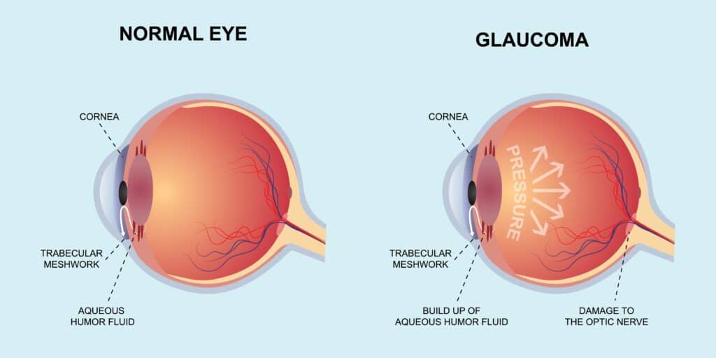
Your eye continuously produces a clear fluid called aqueous humor that circulates inside the eye, nourishing the surrounding tissues. Normally, this fluid drains out through a mesh-like channel near the front of the eye, keeping the pressure inside stable. When that drainage system doesn’t work properly, fluid accumulates and pressure rises.
That elevated pressure is what gradually squeezes and destroys optic nerve fibers over time. The two most common forms of the disease develop differently based on where the drainage problem occurs:
- In open-angle glaucoma, the drainage channel is accessible but not functioning efficiently, so pressure climbs slowly and silently over years
- In narrow-angle glaucoma, the space between the iris and cornea is physically too tight, blocking fluid from escaping. A sudden blockage of this kind can cause a rapid, painful pressure spike that requires immediate attention.
Each type of glaucoma requires a different treatment approach, and getting that distinction right is part of what a proper evaluation at Kirk Eye Center determines.
It is also worth knowing that a small number of cases develop without elevated pressure at all. In these situations, the optic nerve appears to be unusually sensitive, and damage occurs even when pressure readings look normal. This is one reason a full evaluation goes well beyond a simple pressure check.
What Happens to Your Vision When the Optic Nerve Is Damaged?
Glaucoma damages vision gradually. It chips away at the edges first, typically starting with peripheral vision, the area on the outer sides of what you can see. Most people miss this entirely because the brain quietly compensates, filling in the gaps without any obvious signal that something is wrong. As the condition progresses without treatment, that affected area gradually closes in. Side vision narrows, central vision can eventually be pulled into the damage, and in severe, untreated cases, patients are left with only a narrow tunnel of sight before blindness follows.
Unfortunately, vision changes due to glaucoma are irreversible. The nerve fibers that once carried those visual signals are gone for good.
Glaucoma earned its reputation as the “silent thief of sight” because it often causes no pain and no obvious symptoms in its early stages. Your eyes don’t water, your vision doesn’t obviously blur, and your daily routine feels completely normal while the damage accumulates.
Unlike cataracts, where patients actively notice clouding develop over time, glaucoma works entirely beneath the surface of your awareness. By the time peripheral loss becomes undeniable, the disease has usually been progressing for years. That is why comprehensive eye exams are the primary way this condition gets caught before it causes serious harm.
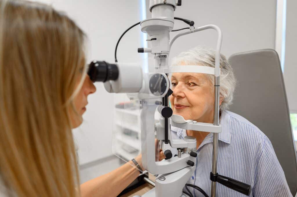
How Glaucoma Is Detected at Kirk Eye Center
A standard vision test will not catch glaucoma. Detecting it requires a specific set of evaluations that go well beyond reading letters on a chart.
At Kirk Eye Center, diagnosing glaucoma starts with measuring the pressure inside your eye, but it doesn’t stop there. Our team also evaluates the thickness of your cornea, examines the drainage angle between the iris and cornea, and maps the nerve fiber layer at the back of your eye using optical coherence tomography, a detailed imaging scan that can detect thinning in the optic nerve tissue before vision loss even begins. Automated visual field testing checks for blind spots you may not have noticed on your own. A careful dilated examination lets our doctors look directly at the health of the optic nerve itself.
Together, these tools give our team a complete picture of what is happening inside your eye, making it possible to identify glaucoma at its earliest and most treatable stage, often long before you would have noticed anything was wrong on your own.
Slowing the Damage Before It Gets Worse
Once glaucoma is diagnosed, the priority shifts to keeping eye pressure under control so the optic nerve is protected from further harm. There are several effective treatment options available: in-office laser procedures, prescription eye drops, and minimally invasive surgery. The right approach depends on the type of glaucoma, how advanced it is, and how the eyes respond to early treatment.

Eye Drops
For many patients, prescription eye drops are an effective first step, used daily to reduce the amount of fluid the eye produces or to help it drain more efficiently.
Laser Treatment
For over a decade, Kirk Eye Center has viewed selective laser trabeculoplasty (SLT) as a first-line option for newly diagnosed glaucoma management. We immediately recognized its benefits over topical drop therapy: continuous action, no dependence on compliance, no serious long-term side effects, repeatability, and efficacy.
Laser treatment for glaucoma is often an excellent option. Selective laser trabeculoplasty, known as SLT, improves the eye’s natural drainage and matches first-line eye drop therapy in effectiveness for the majority of patients. It requires no incisions, has a quick recovery, and causes no damage to surrounding tissue. A newer option called direct selective laser trabeculoplasty, or DSLT, takes a similar approach without the need for a contact lens placed on the eye. The laser is delivered directly through the surface of the eye in just a few seconds, which makes the procedure faster and more comfortable for many patients.
MIGS
For more advanced cases, minimally invasive glaucoma surgery offers a range of targeted options, including micro-sized drainage devices and outpatient procedures that often take under 15 minutes to complete. These options have significantly expanded what is possible for patients who previously had far fewer choices once eye drops stopped working.
Throughout every stage, ongoing glaucoma patient care at Kirk Eye Center means your plan is continuously monitored and adjusted as your condition evolves. Managing glaucoma is a long-term commitment, and the outcome depends heavily on the consistency and quality of the care behind it.
Concerned about your risk for glaucoma or noticing changes in your peripheral vision? Schedule an appointment at Kirk Eye Center at one of our Chicagoland locations.